QUESTION
ANSWER
SKELETON AND MUSCLE
Table of Contents
Introduction
LO1
1.1. Structure of human skeleton and relation of structure to function
Relation of structure to function
1.2. Different types of joint and its importance
1.3. Structure of synovial joint with component parts
1.4. Properties and functions of cartilage, ligaments and tendons
Properties and functions of tendons
Properties and functions of ligaments
LO2
2.1. Properties of three different types of muscles
Skeletal muscle
Smooth muscle
Cardiac muscle
Sliding filament hypothesis
2.2. Extension and flexion of elbow joint by antagonistic muscle
Figure 6: Extension and flexion of elbow joint by antagonistic muscle
LO3
3.1 Effects of bad posture on muscular and skeletal systems
3.2 Effects of poor lifting techniques on muscular and skeletal systems
3.3 Effects of skeletal disease on healthy functioning of skeletal system
Conclusion
Reference list
Introduction
Human skeleton is the internal framework of body composed of different muscles, bones, tendons as well as cartilage. This essay focuses on the structure of human skeleton with clear description of various types of joints. Besides, the properties and functions of different tendons and ligaments are also described here. Along with this, sliding filament hypothesis is taken into consideration for explaining the mechanism of muscle contraction.
LO1
1.1. Structure of human skeleton and relation of structure to function
Structure of human body skeleton is helpful for bipedal locomotion as well as having an erect posture. As opined by Asahara et al. (2017, p.1773), the bony human skeleton is made up of two parts namely axial as well as appendicular skeleton. Components of axial skeleton of central core unit include the skull, vertebrae, ribs and sternum whereas appendicular skeleton comprises of bone extremities.
Skull consists of Cranium, Mandible and Maxilla. Shoulder consists of Clavicle and Scapula. Arm consists of Humerus, radius and Ulna. Hand is composed of Carpal, Metacarpal and Phalanges. Spine consists of Cervical, thoracic, lumber, sacral and coccygeal. Sternum is the prime component of chest. Pelvic girdle consists of Ilium, Ischium and Pubis. Femur, tibia and fibula are primary bones of legs. Ankles are composed of Talus and Calcaneus. Tarsal, metatarsal, Phalanges make the foot structure.
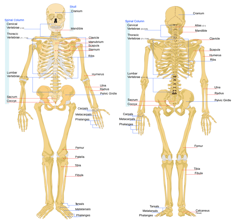
Figure 1: skeletal system and its functions
(Source- askabiologist.asu.edu, 2019)
Relation of structure to function
The bony structure of skeleton is helpful to maintain a specific body shape. As stated by Buenzli and Sims (2015, p.145), the cage like structure of the skeleton helps in protecting the internal organs which is its main function. In addition the hinges and the joints in the skeletal system play a major role in locomotion. It provides a scaffold to the body thus enabling the rest of the body parts to remain in their correct positions. Muscles are an essential component of the structural system of the body. The skeleton supports the muscles and helps in their contraction and relaxation.
1.2. Different types of joint and its importance
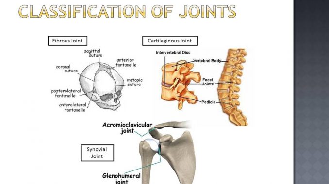
Three main types of joints are present in human body such as fibrous joint, cartilaginous joint and the synovial joint. As mentioned by Golanó et al. (2016, p.945), the fibrous joints are connected with a ligament as occurs in case of tooth which is connected with both radioulnar as well as tibiofibular joints. The cartilaginous joint is seen in between the vertebrae and spine. The synovial joint is highly movable and in elbow and knee it is present leading to the flexion and extension movement.

Figure 2: types of joints in human body
(Source- onlinebiologynotes.com, 2018)
In words of June et al. (2016, p.2050), the fibrous joints are active after birth and help to make the skull bones immovable. Besides, it is helpful to bite and pump blood into dental veins. The cartilaginous joint is allowed to move slightly and act as the ligament to hold the vertebrae together. Moreover, the synovial joint is helpful to absorb shock as well as reduce friction during movement. A smooth layer of hyaline cartilage of bone joint makes the surface slippery.
1.3. Structure of synovial joint with component parts
The three main structure of synovial joint are namely as articular capsule, articular cartilage and the synovial fluid. As described by Katzenberg and Grauer (2018, p.52), the articular capsule consists of two layers such as fibrous as well as synovial layer. White fibre tissue can be found in fibrous layer which helps to hold the articulating bones together. The synovial layer is helpful to mediate nutrition transfer between blood and joint. The articular layer is helpful to minimize the rate of friction during movement. Due to have a smooth cartilage covering it helps to absorb shock.
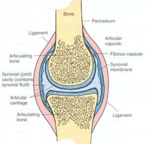
Figure 3: structure of synovial joint
(Source- tutorialpoint.org, 2019)
The synovial fluid is located in the joint cavity performing three vital functions. It basically performs three different tasks such as lubrication of bone joint, nutrient exchange as well as shock absorption (Nourissat et al. 2015, p.225). It is mainly a free movable joint which acts with ligaments.
1.4. Properties and functions of cartilage, ligaments and tendons
Properties and functions of tendons
Tendon is mainly a flexible band of connective tissue that connects the muscles to various bones. It binds to the periosteum of individual’s muscle origin. According to Mungal et al. (2015, p.75), when the muscle contracts tendon takes the lead to transmit the force to the bones which are relatively rigid and hence, pulling of bones causes movement,. Moreover, stretching of tendons is helpful for easy locomotion.
Properties and functions of ligaments
Ligament is mainly a small band which is composed of white, fibrous and elastic tissues. They are helpful to manage the bone action to prevent dislocation. Besides, the elasticity is beneficial to increase length under pressure. As mentioned by Golanó et al. (2016, 950), ligaments are also advantageous to limit some actions such as hyperextension and hyper flexion along with some directional movement of bones.
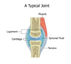
Figure 4: cartilage, ligaments and tendons
(Source- socratic.org, 2019)
It is a flexible connective tissue in nature and resists excessive stretching. Cartilage is found in joint, rib cage, ear as well as throat. In the view of Kjøbsted et al. (2018, p. 1742), it helps to connect bones together and creates places for developing bones. Besides, cartilage helps to reduce the antagonistic functions of bones in various joints. Sometimes damage in cartilage causes damage to tendons and muscles and finally leads to pain.
LO2
2.1. Properties of three different types of muscles
Three types of muscles in human body are named as skeletal, smooth and cardiac muscle.
Skeletal muscle
They are the voluntary muscle that is helpful to control the body movement. It is striated muscle and controlled by somatic nervous system. As described by Martin et al. (2015, p.176), during movement the skeletal bones, muscles and the tendons work together. Major skeletal muscles in the body are located in shoulders, pectorals, and abdominals as well as in quadriceps. Skeletal muscle mainly has four properties. It reacts to nervous stimulation, helps in extensibility and critically involved in maintaining elasticity as well as contractility of muscle.
Smooth muscle
It is the kind of non striated muscle and able to maintain synchronous contractions. It is helpful in maintaining contractility and helpful to transmit mechanical stimuli. Besides, the chemical and electrical stimuli are also transferred through these muscle fibres. The smooth muscle is involved in ATP hydrolysis as well as maintaining phenotypic plasticity by synthesizing collagen, elastin, proteoglycans.
Cardiac muscle
It is an involuntary muscle. As opined by Mungal et al. (2015, p.74), the cardiac muscle is thick as it contracts to move blood in and out of heart. The four basic properties of cardiac muscle are as rhythmicity, contractility, conductivity and excitability. Conduction of impulse from one cell to another is facilitated by conductivity whereas the response to mechanical, electrical and chemical stimuli is performed by excitability of cardiac muscle.
Sliding filament hypothesis
The sliding filament theory of muscle contraction focuses on the generation of force and change of length during muscle contraction. In the process, binding, movement as well as the release of proteins occur. In the view of Buenzli and Sims (2015, p.148), the theory involves four major steps.

Figure 5: Types of muscles
(Source- medlineplus.gov, 2019)
At first the muscle activation is performed by association of myosin with ATP. Thereafter in the second step the breakdown of myosin occurs and cross bridge formation occurs. After that the recharging step includes the pulling stroke which pulls actin to the centre of myosin. The last step involves the cross bridge detachment which allows myosin to bind with ATP. The actions are released and cycle is repeated.
2.2. Extension and flexion of elbow joint by antagonistic muscle
The muscles that are enables elbow flexion are located at the anterior compartment. These mussels are named as biceps brachii, brachialis and brachioradialis. The biceps brachii helps to twist the forearm. It helps in primary movement of the elbow (Katzenberg and Grauer, 2018, p.59). The brachialis and the brachioradialis are the two muscles which are involved in pronation and supination of elbow.
Besides, the triceps and anconeus are the muscles involved in extension of elbow. It has been seen that triceps help to extend the upper arm and stabilization of the elbow. Along with this, the anconeus is also involved in extension of arm at elbow. The triceps become the antagonist muscle as it powers the motion when the biceps lengthen passively to move the elbow.
The same is seen in case of knee movement. As mentioned by Nourissat et al. (2015, p.226), the biceps femoris, semi-membranosus, popliteus and gracilis are the knee muscles that act as antagonist in extension whereas the vastus lateralis, vastus medialis and the rectus femoris are the antagonist muscle for knee reflexion.
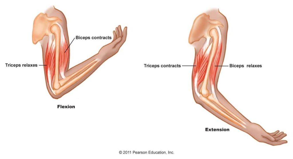
Figure 6: Extension and flexion of elbow joint by antagonistic muscle
(Source- blogs.ncl.ac.uk, 2016)
LO3
3.1 Effects of bad posture on muscular and skeletal systems
Skelton acts as the support system to the entire body. As described by Maruyama et al. (2017, p.47), poor posture may be caused by various physical diseases, infections, nervous system disorders. These kind of bad postures cause serious problems to muscular and skeletal system. The effects of bad posture are described through the following discussion.
Slouching is an abnormal position of the body which makes our muscles to work harder. Hence it creates soreness and pain along with long term health issues. Besides, poor posture affects the foot structure of human body. Poor postures cause carpal tunnel syndrome by affecting the muscles of arms, wrists as well as hands. Muscles become tight in this case. In case of severe effects of poor posture on muscular and skeletal system it causes the past pain to turn into osteoarthritis. In words of Kim et al. (2015, p.1792), prolonged position in slouching causes muscles and tendons to be shorten which causes pain when they are stretched. Thus the poor postures affect our muscular and skeletal system creates pain on spine and lower back.
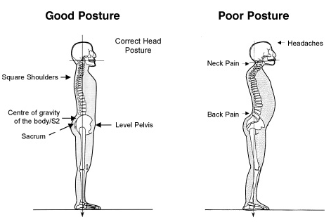
Figure 7: Effects of bad posture on muscular and skeletal systems
(Source: Hypervibe.com, 2019)
3.2 Effects of poor lifting techniques on muscular and skeletal systems
Poor lifting techniques affect greatly on the activities of muscular and skeletal system. According to Chirchir et al. (2017, p.535), poor lifting techniques can cause severe injury on back. Improper lifting techniques cause various types of musculoskeletal disorders with the symptoms of localised pain, dull aches, swelling, stiffness of the body, fatigue and lastly the twitching muscles.
Poor lifting affects the lower back muscles and more stress causes tearing in muscle which is called as muscle strain. In addition, injury in discs is also caused by poor lifting techniques. The skeletal discs in spine can be injured for bulging, breaking into open manner or rupturing when a person lifts a heavy thing in a poor manner. As opined by Kim et al. (2015, p.1793), joint injury is also caused by lifting things following a bad posture. Sometimes it has been seen that poor lifting techniques may cause strains and causes the joints to be locked.
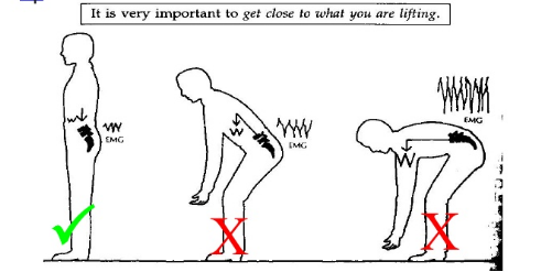
Figure 8: Effects of poor lifting techniques on muscular and skeletal systems
(Source: Influenced by Maruyama et al. 2017, p.50)
3.3 Effects of skeletal disease on healthy functioning of skeletal system
Various types of skeletal diseases and disorders affect the activities of skeletal system. Metabolic bone diseases affect the bone strength as well as their integrity. As mentioned by Chirchir et al. (2017, p.540), the arthritis causes inflammation in bone joints and causes limited range of movement. Thereby, it results into breakdown of cartilage. In this respect, it has been seen that arthritis causes the bone curvature which leads to some kind of disorders such as kyphosis (rounding the upper portion of neck), Lordosis (Rounding the lower back) and lastly the scoliosis (curvature of spine into S shape). Thus, the skeletal diseases affect the proper functioning of musculoskeletal system. Knee functioning is severely affected in this type of bone disorder.
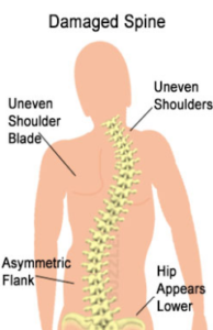
Figure 9: Effects of skeletal deformation
(Source: Influenced by Kim et al. 2015, p.1794)
Conclusion
Thus it can be concluded that the muscles and bones of human skeleton act synergistically or antagonistically to keep the body in full motion. The different types of joints are helpful to maintain the balance between various arms and reduce the rate of friction during movement. The smooth, cardiac and skeletal muscles are essential to maintain balance within a human body. The sliding filament hypothesis focuses on the steps involved in contraction and relaxation of muscles.
Reference list
Asahara, H., Inui, M. and Lotz, M.K., (2017). Tendons and ligaments: connecting developmental biology to musculoskeletal system. Journal of Bone and Mineral Research, 32(9), pp.1773-1782.
askabiologist.asu.edu. (2019). Bone Basics and Bone Anatomy. Available at: https://askabiologist.asu.edu/bone-anatomy [Accessed on: 06/03/2019]
blogs.ncl.ac.uk. (2016). Skeletal muscle structure and function. Available at: https://blogs.ncl.ac.uk/katarzynapirog/files/2016/01/antagonistic_muscle_groups1336591927160.jpg[Accessed on: 06/03/2019
Buenzli, P.R. and Sims, N.A., (2015). Analysing the osteocyte network in the human skeleton. Bone, 75, pp.144-150.
Chirchir, H., Zeininger, A., Nakatsukasa, M., Ketcham, R.A. and Richmond, B.G., (2017). Does trabecular bone structure within the metacarpal heads of primates vary with hand posture?. Comptes Rendus Palevol, 16(5-6), pp.533-544.
Golanó, P., Vega, J., de Leeuw, P.A., Malagelada, F., Manzanares, M.C., Götzens, V. and van Dijk, C.N., (2016). Anatomy of the ankle ligaments: a pictorial essay. Knee Surgery, 24(4), pp.944-956.
Hypervibe.com (2019). Effects of poor posture. Available at: https://www.hypervibe.com/au/blog/not-seeing-results-maybe-youre-training-the-wrong-muscles/ [Accessed on 2nd March 2019]
June, R.K., Liu‐Bryan, R., Long, F. and Griffin, T.M., (2016). Emerging role of metabolic signaling in synovial joint remodeling. Journal of Orthopaedic Research, 34(12), pp.2048-2058.
Katzenberg, M.A. and Grauer, A.L. eds., (2018). Biological anthropology of the human skeleton. UK: John Wiley & Sons.
Kim, D., Cho, M., Park, Y. and Yang, Y., (2015). Effect of an exercise program for posture correction on musculoskeletal pain. Journal of physical therapy science, 27(6), pp.1791-1794.
Kjøbsted, R., Hingst, J.R., Fentz, J., Foretz, M., Sanz, M.N., Pehmøller, C., Shum, M., Marette, A., Mounier, R., Treebak, J.T. and Wojtaszewski, J.F., (2018). AMPK in skeletal muscle function and metabolism. The FASEB Journal, 32(4), pp.1741-1777.
Martin, R.B., Burr, D.B., Sharkey, N.A. and Fyhrie, D.P., (2015). Mechanical properties of ligament and tendon. In Skeletal Tissue Mechanics (pp. 175-225). Springer, New York, NY.
Maruyama, K., Takemura, N., Martino, M.M., Kondo, T. and Akira, S., (2017). Netrins as prophylactic targets in skeletal diseases: A double-edged sword?. Pharmacological research, 122, pp.46-52.
medlineplus.gov (2019). Types of muscle tissue. Available at: https://medlineplus.gov/ency/images/ency/fullsize/19917.jpg[Accessed on: 06/03/2019]
Mungal, S.U., Dube, S.P., Dhole, A., Mane, U. and Bondade, A.K., (2015). New hypothesis for mechanism of sliding filament theory of skeletal muscle contraction. National Journal of Physiology, Pharmacy and Pharmacology, 5(1), pp.72-87.
Nourissat, G., Berenbaum, F. and Duprez, D., (2015). Tendon injury: from biology to tendon repair. Nature Reviews Rheumatology, 11(4), pp.223-233.
onlinebiologynotes.com. (2018). Classification of joints. Available at: https://www.onlinebiologynotes.com/wp-content/uploads/2018/01/classificationof-joints-678×381.jpg [Accessed on: 06/03/2019]
socratic.org. (2019). What is the relationship between ligament, cartilage, muscle and tendon? Available at: https://socratic.org/questions/what-is-the-relationship-between-ligament-cartilage-muscle-and-tendon Properties and functions of cartilage [Accessed on: 06/03/2019]
tutorialpoint.org. (2019). Structure of Synovial Joints. Available at: http://www.tutorialpoint.org/ProvaBiswas/Synovial_joint_structure.jpg [Accessed on: 06/03/2019]
Looking for best Biology Assignment Help. Whatsapp us at +16469488918 or chat with our chat representative showing on lower right corner or order from here. You can also take help from our Live Assignment helper for any exam or live assignment related assistance